Diagnosis of Neuromas: What are the Techniques Used?

Understanding Flat Feet in Kids: What Parents Need to Know
October 17, 2023
7 Things to Consider When Choosing the Best Achilles Tendon Repair Doctor in Houston [Infographic]
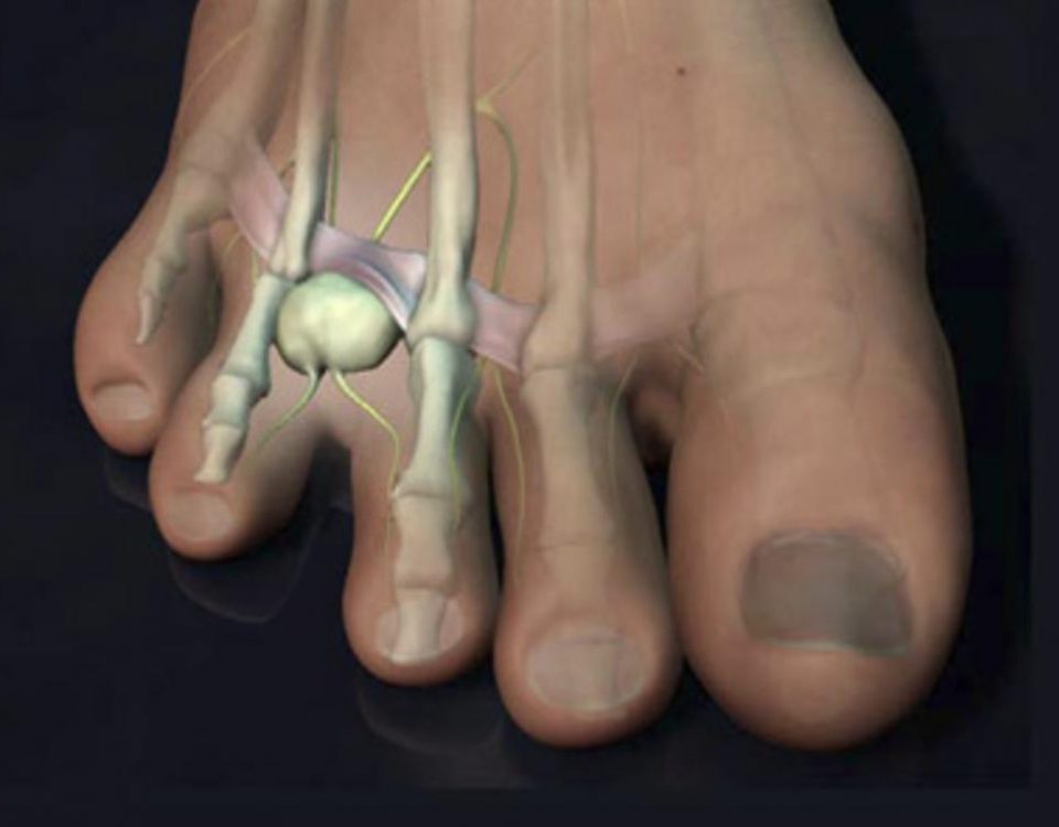
October 30, 2023Understanding the nature and source of neuroma foot pain is a multi-faceted endeavor that calls upon a blend of diagnostic methods. In Houston, medical professionals follow a systematic approach that encompasses the patient’s history, physical examination, and imaging techniques such as ultrasound. This article delves into each of these components in detail to offer insights into how neuroma, a benign nerve tissue tumor often causing pain, is diagnosed.
Gathering Patient History
The initial phase in the diagnostic journey involves an in-depth conversation between the doctor and the patient. When a person consults a specialist for neuroma foot pain in Houston, the medical expert conducts a detailed interview. The objective is to pinpoint common signs and symptoms often associated with neuroma. These can range from pain in the space between two neighboring toes, a tingling or numbing sensation in the toes, discomfort in the ball of the foot, to more unique symptoms like a feeling of walking on pebbles or a bunched-up sock. Patients may also report a burning sensation in their feet or toes, along with periodic jolts that resemble electrical shocks.
Comprehensive Physical Examination
Following a complete history intake, the next step is a meticulous physical examination. Here, the physician scrutinizes various aspects of the foot, paying close attention to vascular, dermatological, neurological, and orthopedic markers. Special focus is given to the area of pain, most commonly situated between the third and fourth toes in the ball of the foot, followed by the second and third toes as the second most frequent site.
The doctor will employ a range of methods, including palpation of the target area by pressing up the ball of the foot between the metatarsal heads. Pressure may also be applied across the forefoot to stimulate the nerve further.
Mulder’s Sign
In addition, a specialized test known as Mulder’s Sign may be executed. This involves a combination of pressure applications from the side and the bottom of the foot simultaneously. Another aspect to be examined is the sensory deficit inside the toes, as neuromas can induce numbness in that particular zone.
Mechanical Tests
Maneuvering the third and fourth metatarsal heads in opposite directions while pressing them together is another mechanical technique frequently utilized for diagnosing neuroma.
Role of X-Rays
It should be noted that X-rays are not particularly effective for directly visualizing neuromas or nerves. However, they are instrumental in eliminating other potential underlying conditions that might mimic neuroma symptoms. This includes stress fractures, metatarsal fractures, joint dislocations, and even changes in the joints due to arthritis.
Utility of Ultrasound
Ultrasound is a widely-used imaging tool for neuroma diagnosis, capable of differentiating between varying tissue densities. It is particularly useful when inflammation or fluid is present in the foot, appearing as a black halo around the inflamed nerve in ultrasound images. The sound waves reflected by the nerve can offer insights into its size and other characteristics, such as whether it is compressible.
Concluding Remarks
Apart from the diagnostic measures discussed, doctors may also opt for diagnostic injections, MRI scans, differential diagnosis, and even use treatments as a diagnostic tool for neuroma identification. These components collectively form the most comprehensive approach for diagnosing neuroma foot pain in Houston.
Featured Image Source: https://media.istockphoto.com/id/1402571496/photo/asian-woman-feeling-pain-in-her-foot-at-home.webp?b=1&s=612×612&w=0&k=20&c=64NlbPr6r_ARh2apL60vtyNgA1ozKbrWJpE93tQU8uQ=


